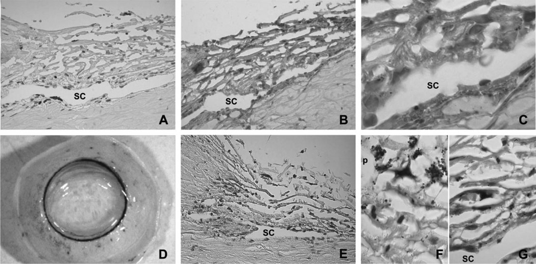
Fig. 5.
Immunohistochemical localization of TGF-β1 in the outflow pathway in human paraffin sections: (A) Negative control using a rabbit non-specific IgG as primary antibody; (B) low magnification of the outflow pathway stained with anti-human TGF-β1 antibody (brown precipitate); and (C) high magnification of the same section showing the presence of TGF-β1 in the ECM surrounding the TM and SC cells. Analysis of the TGF-β1 gene promoter expression in the outflow pathway: (D) Macroscopic observation of a human perfused anterior segment transduced with 107 pfu of AdTGFb1-LacZ showing that positive β-galactosidase staining associated with the expression of TGF-β1 promoter was localized in the outflow pathway (blue staining); (E) paraffin section from the same eye showing that the expression of the TGF-β1 promoter was restricted to some specific cells within the outflow pathway; and (F and G) higher magnification of the TM and SC showing variable levels of LacZ expression in independent cells. Similar results were obtained in six independent experiments. Pigment, p.

