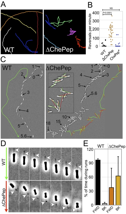
FIG 3 .
ChePep reduces switching of the flagellar rotational direction. (A) Magnified tracings of both WT and ∆ChePep cell swimming behavior (see Movie S1 in the supplemental material). (B) Quantification of reversals per minute of the WT, ∆ChePep, and ChePep* (∆ChePep complemented in trans) strains. (C) DIC video microscopy of WT and ∆ChePep cell swimming (see Movie S2 in the supplemental material). The position of a single swimming bacterium over time is shown for the WT versus the ∆ChePep mutant. The position at a particular time (in seconds) is marked with arrows, and the swimming path is marked in green in one direction and red when the bacteria reverse swimming direction. Inset images of ∆ChePep cell movement at higher magnification are also shown. (D) High-magnification phase-contrast video microscopy shows a ∆ChePep mutant swimming backwards with the flagella “pulling” (see Movie S3 in the supplemental material). The arrows on the left indicate the direction that the bacteria are swimming. (Green indicates swimming forward, and red indicates swimming backward.) The position of the flagella is marked with an arrowhead in each panel. (E) Quantification of the percentage of time that WT and ∆ChePep cells swim forward (FWD) with the flagella “pushing” or backwards (BK) with the flagella “pulling.” n = 8 for each strain. P values are from the two-tailed Student t test.

