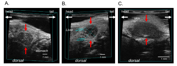
Figure 1.
Representative images of pancreatic tumor margins by ultrasound imaging. Difference in echogenity define the tumor from the surrounding tissue. Tumors are hypoechogen (dark-grayish) compared to surrounding tissue. (A) primary orthotopic pancreatic tumor (red arrows). The light coloured specks (hyperechogenic) within the tumor may be microcalcifications or fatty deposits. (B) The cross-sectional area of the primary pancreatic tumor is depicted. The black areas (anechoic) within the tumor are fluid filled cysts correlating to necrotic areas. (C) Peritoneal tumor with surrounding skin.

