Figure 2.
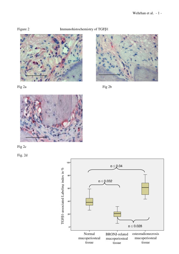
TGFβ1 expression is reduced in BRONJ-related, but increased in osteoradionecrosis-related mucoperiosteal tissue. (a-c) Representative immunohistochemically stained tissue sections show cytoplasmic TGFβ1 staining at × 200 magnification. Scale bars are 100 μm. (a) Immunohistochemical image showing TGFβ1 staining throughout the mucoperiosteal tissue of the jaw. Staining was distributed homogenously throughout the soft tissue. (b) Cytoplasmic staining for TGFβ1 was reduced in BRONJ-related mucoperiosteal tissue accompanied by reduced cellular density. (c) Osteoradionecrosis-related tissue showed higher stained-cell density than normal or BRONJ-related mucoperiosteal tissues. (d) The labeling index for TGFβ1 expression (Table 1) was significantly decreased (p(0.032) in BRONJ-related mucoperiosteal tissue, but significantly increased (p(0.04) in osteoradionecrosis-related tissue, compared to that for normal mucoperiosteal tissue.

