Figure 5.
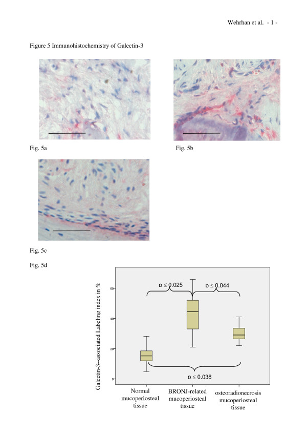
Galectin-3 expression is increased in BRONJ-affected and osteoradionecrosis-related mucoperiosteal tissues. (a-c) Representative immunohistochemically stained tissue sections show cytoplasmic Smad-7 staining at × 200 magnification. Scale bars are 100 μm. (a) Expression of Galectin-3 in healthy mucoperiosteal tissue was restricted to the periosteal margin and cells adjacent to the bone-soft tissue interface. (b) Galectin-3 expression in the BRONJ-affected mucoperiosteal tissue was distributed throughout the entire soft tissue. (c) Osteoradionecrosis-related mucoperiosteal tissue also showed Galectin-3 staining. (d) The relative number (labeling index) of Galectin-3-expressing cells was significantly increased in BRONJ (p(0.025) and osteoradionecrosis samples (p(0.038) compared to control (Table 1).

