
Figure 3. LPS induces inflammatory cytokines and leads to the downregulation of FAO gene expression in cardiac myocytes.
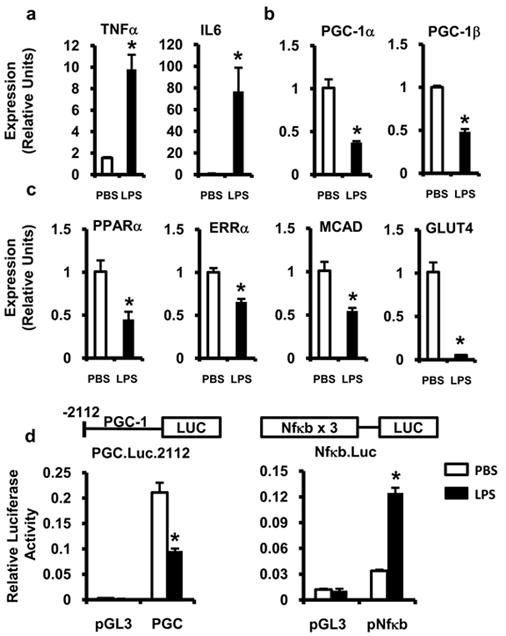
Neonatal rat cardiac myocytes were stimulated with LPS (black bars) or PBS (white bars) for 18h after which gene expression of inflammatory cytokines (a), PGC-1α and β (b), and PGC-1-target metabolic genes (c) were determined by qRT-PCR. All samples were normalized to 36B4. (d) Cardiac myocytes were transfected with a PGC-1α promoter luciferase construct (PGC), NF-κB reporter construct (pNF-κB), or empty vector (pGL3) followed by treatment with LPS (black bars) or PBS (white bars). Relative luciferase activity was determined 18h after stimulation and normalized to SV40 renilla luciferase expression. Experiments were performed in triplicate and reported values represent mean ± SE; * p≤ 0.05 for LPS vs. PBS (Student’s t-test (a-c), two-way ANOVA (d)).