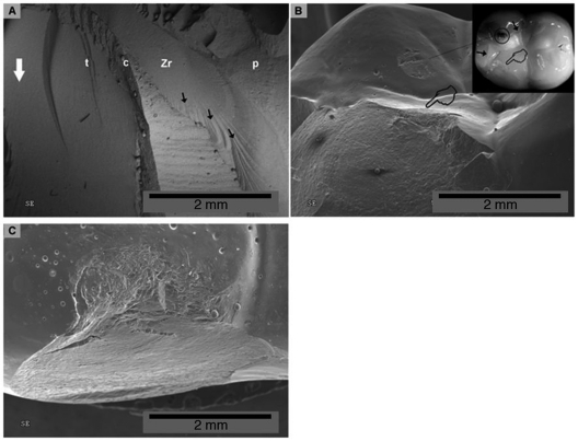
Figure 3.
Fractographic evaluation of failed specimens. (A) Single load-to-fracture: hackles (black arrows) originating from crack initiated on bottom part of the core in a Lava-Espe crown (radial crack). (B) Sliding-contact mouth-motion step-stress fatigue crown: SEM and polarized light microscopy (insert) aspect of indenter damage on lingual cusp (circle), after sliding through lingual slope of buccal cusp and causing a chip-off (pointers). Note that the indenter extended damage to interproximal and lingual cusps by developing cone cracks (black arrows). (C) Most common failure mode was chipping of veneering porcelain not reaching the core/ceramic interface, as depicted in this occlusal view of the buccal cusp. Tooth (t), cement (c), Zr core (Zr), veneering porcelain (p). White arrow points to the cervical margin.

