
Figure 2.
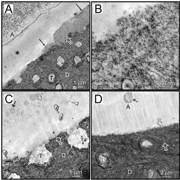
Representative TEM images of 18-month-aged hybrid layers created by water wet-bonding in the absence/presence of CHD and bonding with Scotchbond Multi-Purpose. C, composite; A, adhesive. Between open arrows: hybrid layer. D, dentin; T, dentinal tubule. (A) An example of partial degradation (arrows) of the hybrid layer in the subgroup without CHD. Regions with through-and-through disappearance of the hybrid layer from the composite side to the dentin side (asterisk) were supported by the laboratory-infiltrated epoxy resin. (B) High magnification of the region indicated by arrows in (A) showed loss of fibrillar integrity in the degraded hybrid layer. Pointers: lateral branches of the dentinal tubules. (C) High magnification of the through-and-through region in (A). That space contained denatured collagen components (open arrowhead) and possible microbial invasion (pointers). Arrow: polyalkenoic acid copolymer component of the adhesive. (D) An example of an intact hybrid layer that was sometimes seen in the CHD subgroup bonded by the same adhesive. Arrow: polyalkenoic acid copolymer.