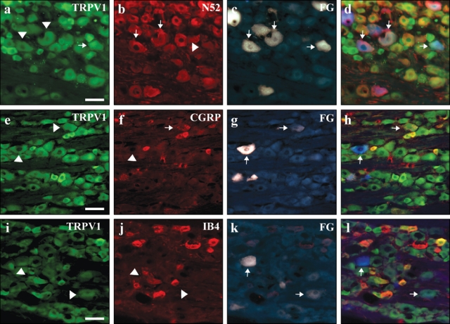
Figure 2.

TRPV1 is infrequently expressed in the cell bodies of neurons innervating the dental pulp. Sections of trigeminal ganglia containing pulpal neurons that were retrogradely labeled with Fluoro-Gold were evaluated for TRPV1 and TRPV2 expression. Tailed arrows point to cell bodies that expressed Fluoro-Gold and/or immunoreactivity for the indicated antigen. Arrowheads highlight cells that expressed Fluoro-Gold and were immunonegative for the indicated antigen. TRPV1 was infrequently expressed in pulpal neurons (Panels a, e, i), but even these few were often NF200-positve (Panel b) and sometimes CGRP-positive (Panel f). The non-peptidergic nociceptive marker IB4 was not found in pulpal neurons (Panel j). All scale bars = 200 µm.
