Figure 4.

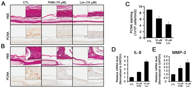
Pamidronate attenuates the re-epithelialization of oral mucosal cells. A 3D oral mucosal wound-healing model was established by the organotypic raft culture system. (A) Five days after air-lifting of the transwell, the oral mucosal tissue constructs were fed with medium only (CTL), medium containing 10 µM PAM, or medium containing 10 µM lovastatin (Lov). The oral tissue constructs were harvested 2 wks after the air-lifting, and subjected to H&E staining (top panel) and IHC staining against IgG (1:200, not shown) and PCNA (1:200) (bottom panel). (B) The circular wound was created 1 wk after the air-lifting. The oral tissue constructs were harvested after 7 days and subjected to H&E staining (top panel) and IHC staining against PCNA (bottom panel). (C) Three independent fields were randomly selected, and PCNA-positive cells were counted per mm2. The bar represents the standard deviation. (D, E) The epithelial cells of the oral mucosal tissue constructs were dissected by LCM. Total RNAs were isolated, cDNAs were synthesized, and qRT-PCR was performed for the expression of IL-8 (D) and MMP-3 (E). The experiments were performed in triplicate, and values were normalized to GAPDH.
