Figure 2.

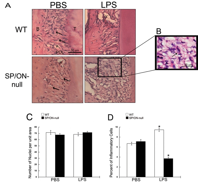
SP/ON-null exhibit disorganized collagen in the PDL and decreased inflammation. (A) Representative images of H&E-stained sections. The PDL in WT mice was highly organized, with fibroblasts tightly surrounded by interstitium (arrows), while the PDL in SP/ON-null mice exhibited less organized collagen ECM, with gaps (arrows). In LPS-injected sites, the PDL of SP/ON-null mice showed substantial disorganization compared with the PDL of WT mice. (B) Arrows indicate examples of cells that were morphologically identified as inflammatory cells in (D). (C) Quantification of nuclei revealed no statistically significant differences in the total number of cells in the PDL of WT and SP/ON-null mice, regardless of injection of PBS or LPS. Error bars = SEM. (D) Quantification of inflammatory cells revealed a significant decrease in inflammatory infiltrate in the LPS-injected PDL of SP/ON-null mice. * indicates p < 0.001 statistical significance of WT LPS vs. SP/ON-null LPS and PBS controls, and SP/ON-null LPS vs. WT LPS and PBS controls, as determined by one-way ANOVA analysis followed by the Tukey test. Error bars = SEM.
