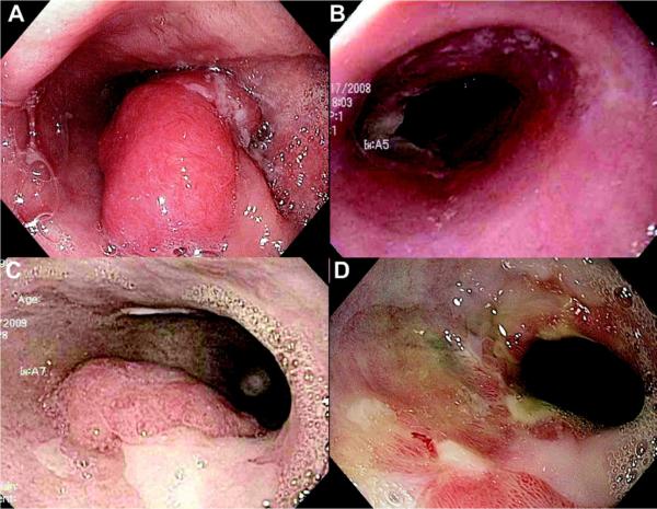
Figure 2.
Endoscopic images of a patient with persistent luminal cancer after concurrent chemotherapy and external beam radiation therapy (chemoradiation) for T2N0 esophageal adenocarcinoma. A, Initial appearance of cancer before chemoradiation. B, One month after chemoradiation, esophagitis but no cancer is detectable. C, Appearance 5 months after chemoradiation, with adenocarcinoma and residual intestinal metaplasia present. D, After 3 cryotherapy sessions, residual cancer is no longer visible. Endoscopic biopsy specimens showed no evidence of cancer. An ink tattoo is visible marking the previous site of the tumor.

