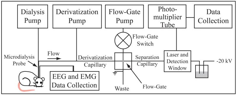
Figure 1.
Schematic of the capillary electrophoresis (CE) instrument. Drawing on the lower left indicates placement of a microdialysis probe in the brain of an awake rat. Dialysate from the probe flows through the derivatization capillary into a flow-gate interface and then is injected onto the separation capillary. Analytes migrate along the separation capillary and through a detection window, where a laser beam induces fluorescence.

