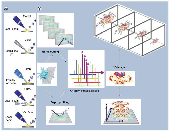
Figure 1. Overall workflow of 3D mass spectral imaging methodology showing (A) ionization method and (B) two types of tissue preparation strategies for mass spectral analysis, serial cutting and depth profiling.
Acquisition of 3D models takes place by collecting mass spectra for each pixel and processing this array into a representative 2D image. 2D images are then stacked in a 3D context to create a 3D model.
DESI: Desorption ESI; LAESI: Laser ablation ESI; LA-FAPA: Laser ablation coupled to a flowing atmospheric pressure afterglow; SIMS: Secondary ion MS.

