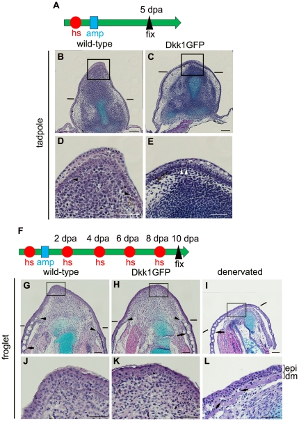
Figure 4. Histological examination of limb blastemas/stumps with or without blockage of Wnt/β-catenin signaling.
(A) Experimental scheme for tadpoles. One heatshock (hs: represented as a red circle) was applied to stage 52 tadpoles 3 to 4 h before amputation. The hindlimb buds were amputated at the presumptive knee level (amp: represented as a blue square), and fixation was done at 5 dpa. (B–E) Longitudinal sections of the limb blastema/stump of wild-type (B, D) and Dkk1GFP (C, E) tadpoles. Panels D and E are high-power views of the boxed regions in B and C, respectively. The distinct basement membrane is indicated by arrowheads. No distinct basement membrane was seen at the tip of the blastema, indicated by arrowheads in (D), while the basement membrane covered the entire amputation plane of the limb stump in (E). (F) Experimental scheme for froglets. One heatshock was applied 3 to 4 h before amputation. The forelimbs of the froglets were amputated through the distal zeugopodium followed by a heatshock every other day, and fixation was done at 10 dpa. (G–L) Longitudinal sections of the limb blastema/stump of wild-type (G, J) and Dkk1GFP (H, K) froglets. Panels J and K are high-power views of the boxed regions in G and H, respectively. Cone-shaped blastemas were formed in both wild-type (G) and Dkk1GFP (H) froglets. These blastemas were covered with dermis-free epidermis, and no skin gland was seen in the blastemal region of wild type (J) or Dkk1GFP froglets (K). In the denervated forelimb, no cone-shaped blastema was formed (I) and a differentiated dermis with skin glands covered the amputation plane at 10 dpa (L). ep, epidermis; dm, dermis. Arrows indicate skin glands. Arrowheads indicate the edge of a distinct basement membrane. Lines indicate the estimated amputation planes. Scale bar = 100 µm for (B), (C), (G), (H), and (I) and 50 µm for (D), (E), (J), (K), and (L).

