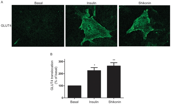
Figure 6. Shikonin stimulates translocation of GLUT4.
(A) Representative confocal image of GLUT4-translocation which was detected by myc-antibody in cells stable transfected with GLUT4myc after 2 h treatment with either 1 µM shikonin or 1 µM insulin (B) Quantification of confocal images obtained in (A) by using Image J and expressed as % of basal. Graph show mean ± SEM of 3 experiments performed. Asterisks represent statistical difference as analyzed by one way ANOVA between basal and treated cells (***p<0.001, *p<0.05).

