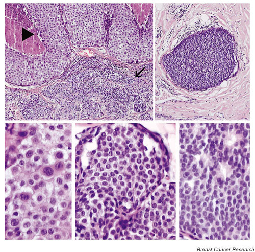
Figure 2.

Differential diagnosis is often difficult between lobular carcinoma in situ (arrow in upper left panel) and low-nuclear-grade, solid ductal carcinoma in situ (upper right panel). Both lesions exhibit characteristic small monomorphic cells with a high nuclear-cytoplasmic ratio (high-power views, lower middle and lower right panels, respectively). In contrast, high-grade ductal carcinoma in situ (arrowhead in upper left panel; high-power view, lower left panel) exhibits markedly different histopathological features, notably the cohesiveness of neoplastic cells, pleomorphic nuclei and abundant eosinophilic-to-amphiphilic cytoplasm. Haematoxylin/eosin stain.
