Abstract
A common trunk of origin of the right testicular and middle suprarenal arteries with a retrocaval course was observed during the dissection of a male cadaver. The Common trunk (CT) arose from the anterior aspect of the abdominal aorta (AA) at the level of the right renal artery (RRA) and after a short course behind the inferior vena cava (IVC), the CT divided into right testicular and middle suprarenal arteries. The middle suprarenal artery (MSA) passed upwards behind the IVC to the right suprarenal gland. The right testicular artery (RTA) descended posterior to the RRA and anterior to the IVC. It then continued on its normal route distally with the right testicular vein. The awareness of such variations of testicular and middle suprarenal arteries and their unusual origin and course might complicate the interpretation of angiograms and surgical procedures in the posterior abdominal area.
Keywords: Right testicular artery, Middle suprarenal artery, Common trunk, Retrocaval
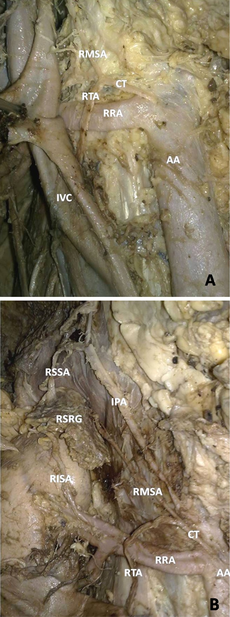
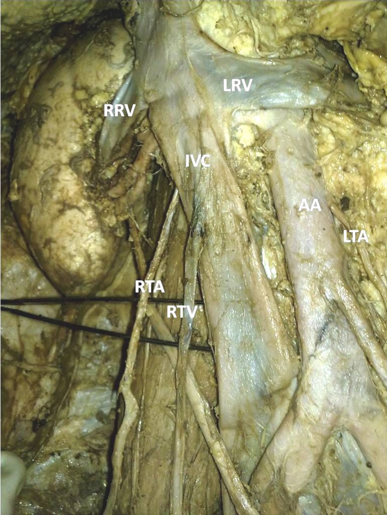
During routine dissection for undergraduate medical students, an abnormal origin and course of the right testicular artery (RTA) and middle supra renal artery (MSA) was detected in a 65-year-old male cadaver. A common trunk arose from the anterior aspect of the abdominal aorta at the level of the right renal artery (RRA). After a short course behind the inferior vena cava (IVC), the common trunk divided into RTA and MSA (Fig. 1 (A and B)). Thereafter, the RTA descended behind the IVC and coursed along its normal route distally accompanied by the right testicular vein (Fig. 2). The left testicular artery and the testicular veins followed the usual description in the same individual. The MSA coursed upwards behind the IVC to reach the right suprarenal gland. The other suprarenal arteries on both sides showed the usual origin and course.
Fig. 1.
a Photograph showing right testicular and middle suprarenal arteries from the common trunk; b Photograph showing supra renal arteries of the right suprarenal gland RTA Right testicular artery; AA Abdominal aorta; CT Common trunk; RRA Right renal artery; IPA Inferior phrenic artery; RSSA Right superior suprarenal artery; RMSA Right middle suprarenal artery; RISA Right inferior suprarenal artery; RSRG Right suprarenal gland
Fig. 2.
Photograph showing retrocaval course of the RTA RTA Right testicular artery; RTV Right testicular vein; IVC Inferior vena cava; LRV Left renal vein; RRV Right renal vein; AA Abdominal aorta; LTA Left testicular artery
Variations of number, origin, and course of the gonadal and suprarenal arteries have been reported [1–4]. Notkovitch (1956) described three variations of the gonadal arteries: (1) where the artery descends directly without any contact with the renal vein, (2) where the artery arises at a higher level than the renal vein and crosses the vein lying directly in front, and (3) where the artery arises at a lower level and arches over the renal vein to descend [2]. This case is similar to Notkovich's type I pattern but with an unusual course behind the vena cava.
Variations in the origin, course, and branches of the testicular arteries can be attributed to their embryological origin. The lateral splanchnic branches of the dorsal aorta give rise to one gonadal and three suprarenal arteries on each side. Those branches that form the testicular arteries enter the mesonephros crossing ventral to the supracardinal vein and dorsal to the subcardinal vein. The lateral splanchnic artery that persists as the RTA normally passes anterior to the supracardinal anastomosis, which, in turn, gives rise to part of the IVC. The coursing of the lateral splanchnic artery posterior to this anastomosis in the embryo may explain why the RTA was seen posterior to the IVC [5].
References
- 1.Adachi B (1928) Das Arteriensystem der Japaner. II(88):73–74.
- 2.Notkovich H. Variations of the testicular and ovarian arteries in relation to the renal pedicle. Surg Gynecol Obstet. 1956;103(4):487–495. [PubMed] [Google Scholar]
- 3.Gagnon R. Middle suprarenal arteries in man: a statistical study of 200 human adrenal glands. Rev Can Biol. 1964;23:461–467. [PubMed] [Google Scholar]
- 4.Bergman RA, Afifi AK, Miyauchi R (1989) http://www.anatomyatlases.org/AnatomicVariants/Cardiovascular/Text/Arteries/Suprarenal.shtml
- 5.Felix W. Mesonephric arteries (aa. mesonephricae) In: Kiebel F, Mall FP, editors. Manual of human embryology vol 2. Philadelphia: Lippincott; 1912. pp. 820–825. [Google Scholar]


