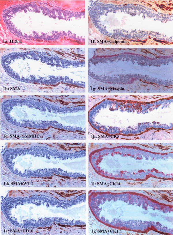
Figure 1.

Immunostaining pattern of ME cells in columnar hyperplasia (case 1). (a) H&E staining; (b) immunostaining for SMA; (c-f) double immunostaining of SMA with SM-MHC, WT-1, CD10, and calponin, respectively, and the segment of the ME layer is negative for all antibodies; (g-j) double immunostaining of SMA with maspin, CK5, CK14, and CK17, respectively, and the segment of the ME layer is positive for all antibodies.
