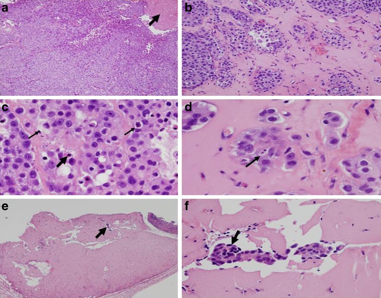
Fig. 5.
Histological changes of C4-2B tumor tissues of mice treated with ethanol extract of neem leaves (EENL; 100 μg/kg body weight). At the end of 11 weeks, xenograft tumor tissue was collected from the mice and stained with hematoxylin and eosin. Two sections of tumor tissue from each mouse and six mice in a group were examined for histological changes. a Control tumor tissue from vehicle-treated mice shows dense tumor cells and the arrow points to the area of coagulative tumor necrosis at ×100. b–f Depicts tumor tissues from mice treated with EENL. b Tumor tissue shows nests of tumor cells separated by hyalinized connective tissue indicated by arrow at 100×, indication of treatment effect. c Tumor hyalinization showing apoptosis, the thin and thick arrows indicate apoptotic bodies and pyknotic nucleus undergoing cell death at ×400. d Tumor hyalinization showing apoptotic nucleus at ×600. e Tumor tissue with hyalinized fibrosis, arrow indicates residual tumor at ×100. f Hyalinized fibrous tissue showing residual tumor cells at ×200 magnification

