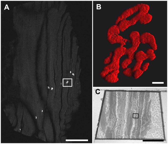
Figure 4.
A. Confocal tile scan of a 10 μm cryostat section of sucrose-protected rat muscle labeled with α-bungarotoxin-AlexaFlour™ 555. The boxed region was targeted for TEM. Scale bar = 500 μm. B. A high magnification three dimensional shadow projection rendering of the boxed NMJ in Fig. 4A. Scale bar= 5 μm. Movies of three-dimensional rendering (Fig. 4B-3Dmovie) and original confocal z-stack (Fig. 4B-Zmovie) available as supplementary data. C. Image of the resin block face. The boxed region corresponds to that in Fig. 4A. Scale bar= 500 μm.

