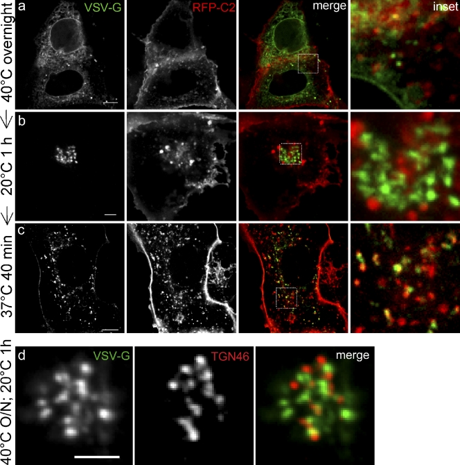
Figure 8.
PS becomes cytosolically exposed during progression through the secretory pathway. COS7 were cells double-transfected with VSVG-GFP and mRFP-Lact-C2 (a–c), then incubated as indicated to label the ER (40°C overnight), the Golgi complex (20°C for 1 h after 40°C overnight), and post-Golgi compartments (37°C for 40 min, after 40°C overnight followed by 1 h at 20°C). Insets show enlargement of indicated merged image. Cells expressing VSVG-GFP (d) were grown overnight at 40°C, then shifted to 20°C for 1 h, fixed, and immunostained for TGN46. Insets show enlarged views of the boxed region in the merged image. Bars, 5 µm.

