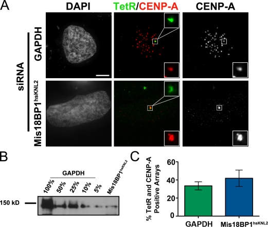
Figure 7.
The Mis18 complex is not required for CENP-A deposition at the LacO/TRE array. (A) Representative images of endogenous CENP-A recruitment in U2OS-LacO cells treated with 15 mM IPTG after 72 h of either GAPDH or Mis18BP1hsKNL2 siRNA treatment. Cells had been transiently transfected with LacI-HJURPScm3 and GFP-TetR 48 h before fixation. Cells were transfected after an initial 24 h siRNA treatment to ensure Mis18BP1hsKNL2 depletion before CENP-A establishment at the array. Insets show magnified views of boxed regions. Bar, 5 µm. (B) Cellular extracts from GAPDH and Mis18BP1hsKNL2 siRNA-treated cells were analyzed by Western blotting using an anti-Mis18BP1hsKNL2 antibody. Each lane contains lysate from 107 cells. (C) Quantification of CENP-A staining at the LacO/TRE array marked by GFP-TetR after 72 h of GAPDH or Mis18BP1hsKNL2 siRNA treatment and 1 h of 15 mM IPTG treatment. At least 30 cells per condition were analyzed; n = 2. Error bars represent the standard deviation between the two experiments. The p-value between GAPDH and Mis18BP1hsKNL2 is 0.3609.

