Figure 9.
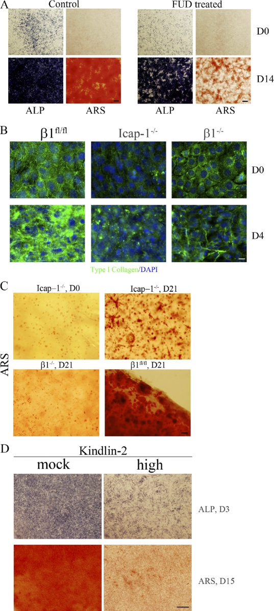
Blocking fibronectin fibrillogenesis impairs mineralization. (A) Wild-type cells were induced to differentiate into osteoblasts in the presence (FUD treated) or absence (control) of FUD, and the expressions of alkaline phosphatase (ALP) and mineralization (Alizarin red S [ARS]) were monitored at day 0 (D0) and day 14 (D14). (B) Wild-type (β1fl/fl), Icap-1−/−, and β1−/− cells were cultured as described for in vitro mineralization assay. At day 0, the medium was changed to induce differentiation. Cells were fixed either at day 0 or day 4, and type I collagen deposition was analyzed by immunofluorescence staining. (C) Wild-type (β1fl/fl), Icap-1−/−, and β1−/− cells were embedded in highly concentrated type I collagen gel (5 mg/ml). After 1 wk in normal medium to allow cell proliferation, the medium was changed for the osteogenic medium, and the culture was continued for an additional 21 d. Gels were then stained with Alizarin red S to detect mineralized foci. (D) Mineralization of cells expressing high levels of kindlin-2 (high) was analyzed after their culture in osteoblast differentiation media. Expression of alkaline phosphatase was used to follow the early commitment of cells to the osteoblast lineage at day 3 (D3), and mineralization was visualized by Alizarin red S staining at day 15 (D15). Bars: (A, C, and D) 1 mm; (B) 20 µm.

