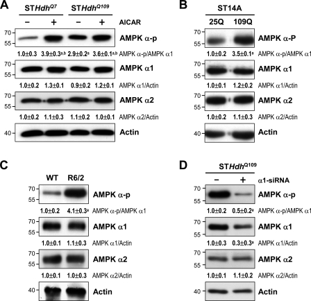
Figure 2.
Expression of mHtt activates AMPK in striatal cells. (A–D) The total lysates were assessed by Western blot analyses. Results were normalized to those of actin. (A) Cells were incubated with or without 1 mM AICAR for 24 h. a, P < 0.05 versus untreated STHdhQ7; b, P < 0.05 between cells treated with and without AICAR. (B) ST14 cells were transfected with the indicated construct (pcDNA3.1-Htt-[Q]25-hrGFP, 25Q; pcDNA3.1-Htt-[Q]109-hrGFP, 109Q) for 48 h. a, P < 0.05 versus untreated 25Q cells. (C) Striatal lysates of 12-wk-old mice were analyzed. a, P < 0.05 versus WT. (D) STHdhQ109 cells were transfected with small hairpin RNA of AMPK-α1 for 72 h. a, P < 0.05 versus control STHdhQ109 cells. Data are presented as the mean ± SEM of three independent experiments. Molecular mass is indicated in kilodaltons.

