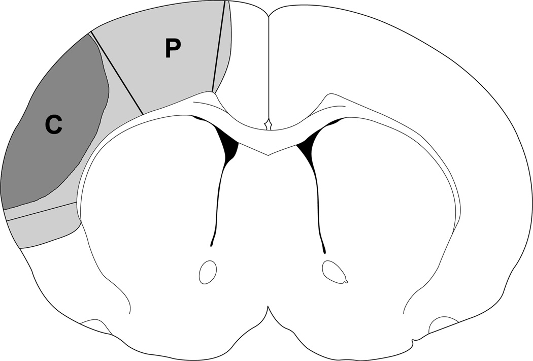
Fig. 1.
A diagram indicates that tissues dissected correspond to the ischemic penumbra and core. Region P plus region C represents ischemic injury in a rat with control ischemia; region P is spared by rapid preconditioning, which is defined as the penumbra, and region C is defined as the ischemic core. These regions were dissected for Western blotting. The corresponding non-ischemic cortex from sham animal without ischemia was dissected for comparison.

