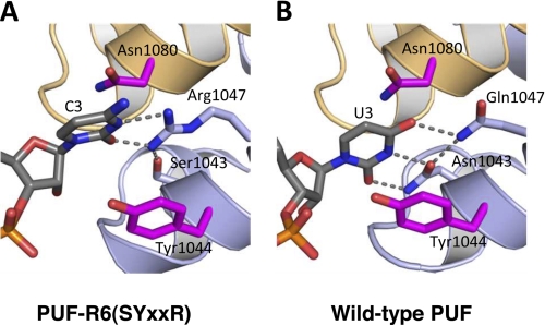
FIGURE 4.
Crystal structure of PUF-R6(SYXXR) in complex with C3 RNA. A, interaction of PUF-R6(SYXXR) with C3 RNA. A ribbon diagram of interaction of repeat 6 with C3 base (complex 1 with chain A and C displayed) is shown. B, interaction of wild-type PUF (NYXXQ) with U3 RNA. A ribbon diagram of interaction of repeat 6 with U3 base is shown. RNA and base-interacting side chains are shown as stick models colored by atom type (red, oxygen; blue, nitrogen; orange, phosphorus). Carbon atoms are colored gray in RNA and light blue in RNA edge-interacting side chains, and magenta inside chains are in position to stack with the RNA base. Hydrogen bonds are indicated with dashed lines. This figure was created with PyMOL.

