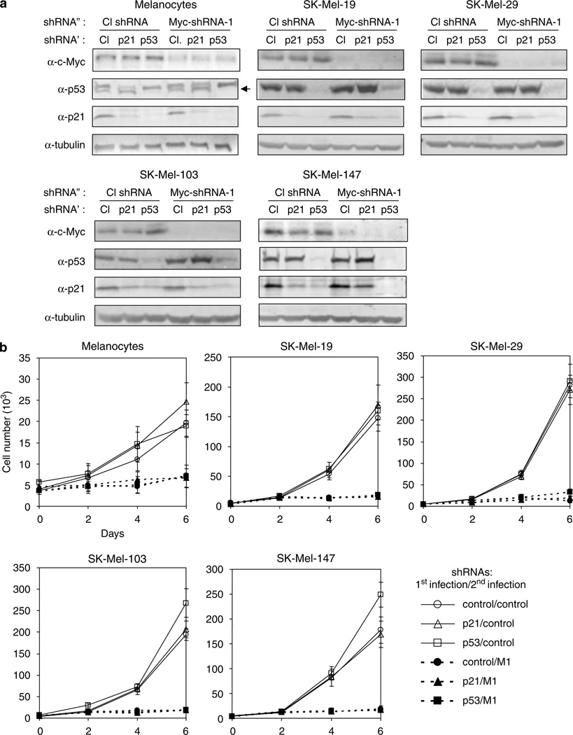
Figure 3.
Inhibition of p53 or p21CIP/WAF does not compensate for the depletion of c-Myc. Cells from the indicated lines were infected with lentiviral vectors expressing control − p53 −or p21CIP/WAF-shRNAs as described in Materials and methods. Two days later, cells were superinfected with control or c-Myc shRNA (M1). (a) Cells were harvested 4days after the second infection and examined by western blotting with the antibodies designated on the right. ShRNA′ and shRNA″ designate shRNAs used for primary and secondary infections, respectively. Note a background band in melanocyte cell extracts probed with p53 antibodies closely migrating with the correct band, designated with the arrow. (b) Cells were plated into 12-well plates and counted daily in triplicates in the presence of trypan blue.

