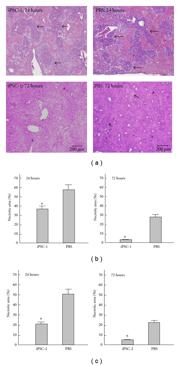
Figure 6.

Effects of iPS cells on histopathological changes in recipient mice. Results showed the representative H&E stain of TAA-treated liver tissue receiving iPSCs or PBS treatment after 24 hours or 72 hours in iPSC clone 1 from our lab and clone 2 from Shinya Yamanaka. In (a) and (b), the necrotic areas in iPSC-treated group were significantly reduced than PBS-treated group in 24 hours and 72 hours after TAA administration in iPSC clone 1. In (c), similar results were identified in iPSC clone 2. *P < 0.05 versus PBS.
