
Figure 1. Double fluorescent in situ hybridization of prdm1a with pax3 and olig4.
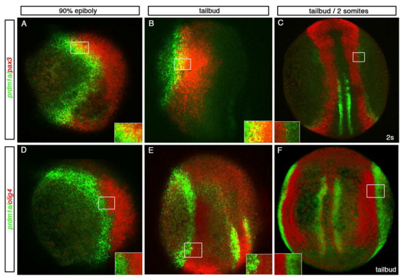
Dorsal and lateral views of 90% epiboly, tailbud and 2 somite stage (9 hpf-11 hpf) confocal micrographs in a single z-stack unless otherwise noted. Insets show higher resolution confocal images for each boxed area. (A-C) prdm1a in green and pax3 in red and (D-F) prdm1a in green and olig4 in red. A lateral view, dorsal to the right, of an embryo at the end of gastrulation (90% epiboly) and lateral view at tailbud exhibit overlap between prdm1a and pax3 (A, B; yellow). By 11hpf, dorsal view of a 2 somite stage embryo, projected image shows the domains are distinct (C). Even at the earliest stages examined, olig4 is distinct from prdm1a. Lateral view of 90% epiboly and dorsal lateral view of a tailbud stage embryo show no overlap in expression (D, E). At tailbud stages shown in a dorsal projected view (F), no overlap is seen in the dorsal domain or the medial adaxial domain.