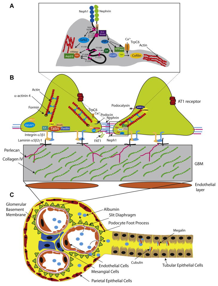
Figure 2. Glomerular filtration barrier and tubules.
A. Nephrin/Neph1 tyrosine phosphorylation dependent recruitment of protein complexes involved in actin regulation. Fyn mediated tyrosine phosphorylation of Neph1 on Y637 and Y638 results in recruitment of adaptor protein Grb2. Similarly nephrin phosphorylation on its tyrosine residues Y1191, Y1208 and Y1232 recruits Nck, Crk (Y1198 and Y1225) and P85 subunit (Y1128, Y1153 and Y1154) of PI3 kinase. B. Schematic cross section of podocyte foot process, glomerular basement membrane and endothelial cells. Also illustrated are the important proteins (mutations or deletions) which have been identified to result in proteinuria in human diseases or mouse models. C. Schematic of the nephron with the proximal tubule illustrating tubular proteins cubulin and megalin. Abbreviations: TrpC6, transient receptor potential C6; nWasp: neural Wiskott Aldrich syndrome protein; Arp2/3: actin related protein 2 and 3; AT1 receptor: Angiotensin receptor 1; SSH1: slingshot 1; PPase: phosphatase.

