
Figure 1.
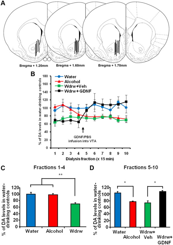
Intra-VTA infusion of GDNF reverses alcohol withdrawal-associated NAc DA deficiency. Rats were trained to achieve a stable level of alcohol intake using the intermittent access to 20% alcohol in two-bottle choice procedure. For the alcohol group (red), microdialysis was performed immediately after the last session of 24 h alcohol access, whereas for the withdrawal group (wdrw) (green and black), microdialysis was performed 24 h after the last drinking session was terminated. An alcohol-naive control group (water) (blue) was included as a control. Samples were collected every 15 min. After collecting four samples, the wdrw group was infused with GDNF (10 μg/μl/side; black) or vehicle (green) into the VTA and returned to the microdialysis chambers for an additional 90 min. A, Schematic representation of the microdialysis probe placement in coronal sections (Paxinos and Watson, 1998). The locations of the dialysis membrane are represented by vertical bars. The numbers indicate the distance anterior to bregma (in millimeters). B–D, NAc DA levels are presented as mean percentage (±SEM) of the baseline of water controls. B, DA levels in the NAc throughout 10 fractions of 15 min in the water, alcohol, wdrw+veh, and wdrw+GDNF groups. C, Average NAc DA levels during fractions 1–4 in the water, alcohol, and wdrw groups. **p < 0.01. D, Average NAc DA levels during fractions 5–10 (following intra-VTA GDNF or vehicle infusion to wdrw groups) in the water, alcohol, wdrw+veh, and wdrw+GDNF groups. *p < 0.05. n = 4–6.