
Figure 3.
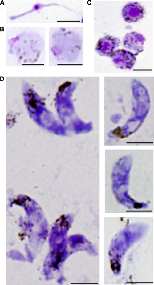
Giemsa-stained thin blood smears of in vitro generated Plasmodium vivax sexual stage parasites and Giemsa-stained thin blood smears of P. vivax sexual stage forms. A, Microgamete with eosinophilic nucleus. B, Round forms with basophilic cytoplasm, dispersed hemozoin, and a single small nucleus not contained within an erythrocyte. C, Round forms with basophilic cytoplasm, dispersed hemozoin, and a large eosinophilic nucleus not contained within an erythrocyte. D, Ookinetes with characteristic elongated form, localized hemozoin, and eosinophilic nuclei. Occasionally, two eosinophilic nuclei are visualized in a single ookinete. Additionally, parasites are seen with one to multiple, occasionally peri-nuclear, foci that do not stain well with Giemsa (→). Scale bars = 5 μm.