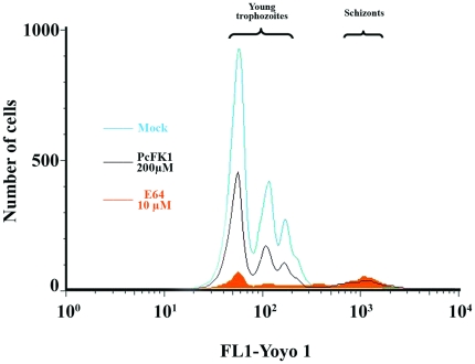
Figure 9. Analysis of merozoites egress/invasion steps on P. falciparum in vitro culture.
In vitro synchronized culture of P. falciparum composed of 0.5% of segmented schizonts was incubated for 12 hr in serum-containing medium and schizonts transition to newly formed trophozoites was analyzed by flow cytometry. Gates were converted to two-dimensional plots illustrating the expected merozoites egress defect in presence of 10  M E64 [51], but also in presence of 200
  M of PcFK1. 91.6% and 56.2% of inhibition of newly formed trophozoites and equivalent segmented schizont accumulation were observed with 10
  M E64 and 200
  M PcFK1 respectively, when compared to the mock control. Mock condition corresponds to a classical P. falciparum culture in presence of 2% DMSO, the vehicle of PcFK1.

