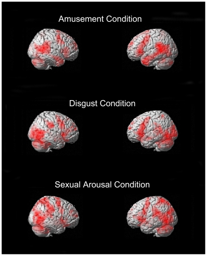
Figure 2. Three-dimensional rendered images of areas of supra-threshold areas of activation for the same contrasts as Figure 1.
Here again, there similarities are obvious between contrasts. All contrasts clearly show involvement of the frontal operculum, dorsolateral prefrontal cortex, premotor and motor cortices, temporo-occipital regions, and cerebellum.

