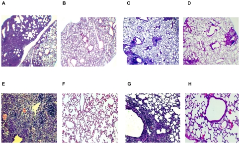
Figure 4. Lung histopathology induced by PR8 infection.
(A and E) Lung resected from an untreated control mouse (Fig. 1) 19 days post-PR8 challenge. (B and F) Lung resected from a normal Balb/c mouse as a control. (C and G) Lung resected from an AdE/in/-2 mouse (Fig. 1) 19 days post-PR8 challenge; each section is a representative of three mice. (D and H) Lung resected from an AdNC/in/-2 mouse (Fig. 1) 19 days post-PR8 challenge; each section is a representative of three mice. Lung sections were examined on a Zeiss Axioskop2 plus microscope using a 2X (A–D) or a 10X (E–H) objective lens in conjunction with an Axiocam digital camera.

