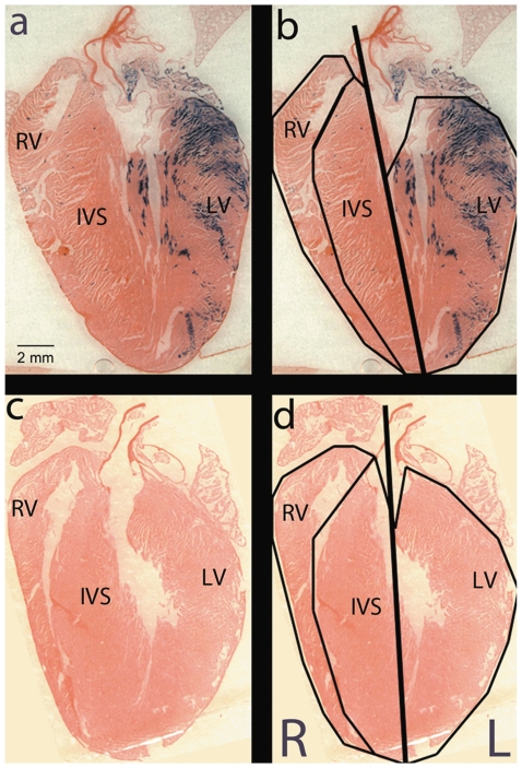
Figure 2. To quantify Pnmt expression, Pnmt +/Cre, ROSA26 +/βgal adult mouse heart images were partitioned into right and left heart.
The line of separation originates from the apex and extends toward the base of the heart and through the aorta (bold line). Additional partitioning isolates the IVS from the RV and LV free walls (panel b and d). Each partition was then analyzed to determine the total pixels containing the blue XGAL+ stain. The left heart shows heavy XGAL+ staining when compared to the right side of the heart. Pnmt+/+, ROSA26βgal/βgal control heart shows no XGAL+ staining (panel c and d).

