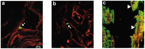
Figure 9. High-magnification confocal co-immunofluorescent staining for Pnmt and sarcomeric α-actinin in adult mouse heart (LV) sections.
Pnmt immunofluorescent staining in the adult mouse heart identifies small triangular-shaped cells (green) (a, b arrows) within the interstitial spaces. Cardiomyocyte-specific staining for sarcomeric α-actinin (red) shows identifiable striations in branching cells. Immunofluorescent co-staining with Pmnt and sarcomeric α-actinin shows apparent Pmnt expression within some cardiomyocytes (c, arrowheads).

