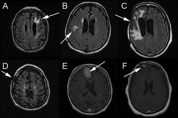
FIGURE 1.
MRI scans highlighting major abnormalities in six patients with OS resulting from lesions are summarized below. Fluid attenuated inversion recovery images are shown in A–D, and gadolinium enhanced T1 images are shown in E and F:
(A) Chronic infarct is in the left frontal lobe. Small cortical infarcts are present in the left frontal lobe. Thalamic chronic lacunar infarcts present.
(B) There is a white matter hyperintensity within both frontal lobes. There is a right internal capsule infarct.
(C) Encephalomalacia in the right frontal and temporal lobes, beneath the right craniectomy at the site of the prior subdural and intraparenchymal hemorrhages. There is associated ex vacuo dilatation of the right lateral ventricle with slight midline shift.
(D) Post-surgical changes in the right calvaria compatible with previous subdural hematoma evacuation.
(E) Meningioma within the right side of the interhemispheric fissure. Old lacunar infarcts. Old right occipital lobe infarct.
(F) Right frontal meningioma.

