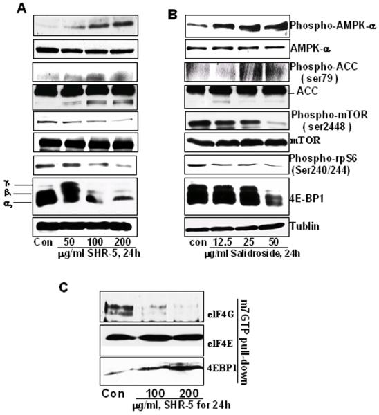
Figure 4.
The effects of SHR-5 and salidroside on AMPKα, mTOR and protein translation initiation in UMUC-3 cells. UMUC-3 cells were treated with vehicle control or indicated doses of SHR-5 or salidroside for 24 hours. At the end of each treatment time, cell lysates were prepared as mentioned in Materials and Methods. A & B: Western blotting analysis of phospho-AMPKα, AMPKα, phosphor-ACC, ACC, mTOR, phospho-mTOR, Phospho-rpS6, and 4E-BP1, and membranes were stripped for reprobing with anti-tubulin antibody for protein loading correction. A representative blot was shown from three independent experiments. C: eIF4E was purified from cell extracts by m7GTP affinity chromatography and probed with antibodies against eIF4E, eIF4G and 4E-BP1. A representative blot was shown from three independent experiments.

