
Fig. 6.
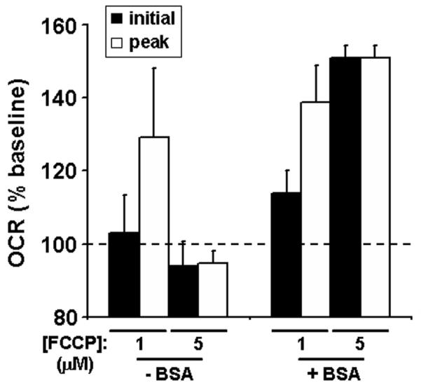
The effect of BSA on FCCP-stimulated O2 consumption rates (OCR). Baseline-normalized initial and peak OCR of organotypic mouse hippocampal slices exposed to FCCP (1 or 5 μM) in the absence (n=4) or presence (n=3) of BSA (4 mg/ml). Initial OCR (black bars) and peak OCR (white bars) were the first or the first or second measurement after FCCP addition, respectively. Data are expressed as mean ± SE and are normalized to the last OCR before FCCP addition. Two slices with baseline OCR below 50 pmol O2/min were excluded from analysis.