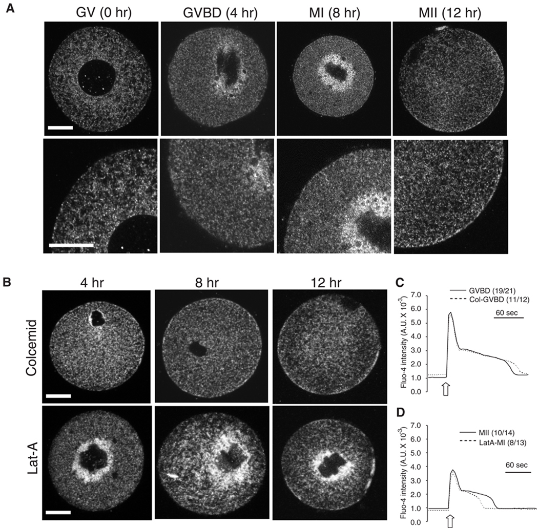
Fig. 7.
Redistribution of IP3R1 during oocyte maturation. (A and B) Oocytes were labeled with IP3R1 and imaged by confocal microcopy. The observations were performed at 0, 4, 8 and 12 hr of maturation, which corresponded with GV, GVBD, MI and MII stages. Oocytes were matured in the absence (A) or in the presence of 5 µM colcemid (B; upper panel) or 2.5 µM Lat-A (B; lower panel). A typical equatorial section is shown. Scale bar, 30 µm. (C) IP3-induced Ca2+ release using cIP3 was examined in control (solid line) and colcemid-treaded (dashed line) GVBD oocytes. The 0.001 sec UV pulse was applied to oocytes (open arrow). The number of oocytes responding to cIP3 is shown in parentheses. (D) Oocytes were matured for 8 hr in the absence of LatA, cIP3 was injected at this time, after which maturation continued for 4 hr in the absence or presence of LatA. IP3-induced Ca2+ release using cIP3 was examined in control MII (solid line) and LatA-treaded MI arrested (dashed line) oocytes. The 0.001 sec UV pulse was applied to oocytes (open arrow). The number of oocytes responding to cIP3 is shown in parentheses.

