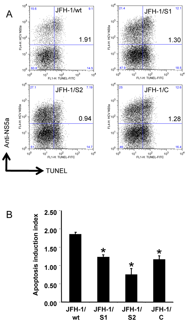
Fig. 4. Apoptosis induction in Huh-7.5.1 cells transfected with JFH-1/wt and its variants.
(A) Three million cells were transfected with 3 µg in vitro transcribed full-genome RNA of JFH-1/wt, S1, S2, and C. Forty-eight hours later, apoptosis was induced by exposing cells to 20 ng/mL TNF-α plus 50 ng/mL Act D. Cells were harvested after 48 h of treatment, and subjected to TUNEL and anti-HCV NS5a staining. Dot-plots show HCV replication and apoptosis at the single cell level. Quadrant gates were determined using unstained- and a TdT-untreated-control in each culture condition. The clone names and apoptosis induction indexes are indicated in the upper right box. (B) Apoptosis induction indexes of JFH-1/wt-, S1-, S2-, and C- transfected cells. Means ± standard deviations of 3 independent experiments are shown. *p < 0.005 compared to JFH-1/wt.

