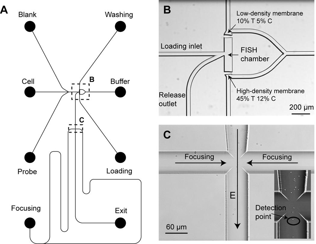
Figure 1.
Schematic of the microchip design for fluorescence in situ hybridization (FISH) and flow cytometry (µFlowFISH). (A) The mask design of the µFlowFISH chip. (B) An image of the FISH chamber formed by two photopolymerized membrane in the channel. (C) The cross-channel structure for electrokinetically focusing microbial cells into a single stream line along the center of the vertical channel for flow cytometry. The enlarged image of the channel cross shows Escherichia coli being focused in the center of the channels.

