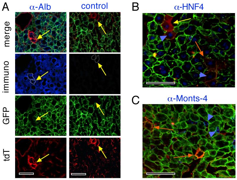
Fig. 3. Alb and HNF-4 expression, but not Monts-4 expression, in “reddish hepatocytes”.
Four days following 2/3 hepatectomy of ROSAmT-mG/+;albCre1 mice, animals were sacrificed, perfused with saline to flush serum albumin from the hepatic circulation, and livers were harvested. (A) Cryosections were immunostained for Alb using anti-mouse Alb antibody (α-Alb, left column of images) or no primary antibody (control, right column of images) followed by Alexa Fluor-350- (blue-) labeled secondary antibody. Slides were mounted without DAPI and were photographed by fluorescence microscopy. Columns of images represent the same frames photographed with the color channels indicated at left. “Reddish hepatocytes” (yellow arrows) in each frame are circumscribed with a fine white line in the Alb images. Yellow arrows are in the same position in each frame of each column of images. (B, C) merged fluoromicrographs of cryosections as in A stained for HNF-4 (B) or Monts-4 (C). Yellow and orange arrows indicate hepatocytes with less (younger) or more (older) green in membranes, respectively. Blue arrowheads indicate representative nuclei in reddish hepatocytes that stained blue for HNF-4 (panel B) or representative Kupffer cells that stained blue for Monts-4 (panel C). Scale bars 100 μmeters.

