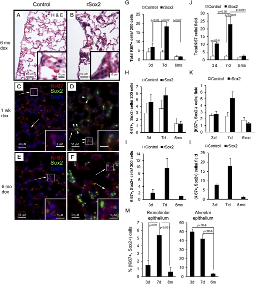
Figure 2.
Exogenous Sox2 caused cell proliferation and epithelial hyperplasia in the respiratory epithelium. Four-week-old mice were treated with doxycyline for the times shown and then killed. (A, B) Hematoxylin-eosin staining of lung sections from mice treated with doxycycline for 6 months demonstrated hyperplastic clusters in the alveolar region of rSox2 mice. (C, D) Dual immunofluorescence for Sox2 and the proliferation marker Ki-67 was used to analyze the effect of transgenic Sox2 expression on proliferation; the intensity of Sox2 fluorescence in rSox2 mice was adjusted to visualize only transgenic Sox2 (Sox2tg). Proliferative cells (Ki-67+) with Sox2tg (arrowheads) or without Sox2tg (arrows) are shown (C–F); pale orange autofluorescence is derived from red blood cells. Three days of doxycyline treatment induced alveolar proliferation (J), and 7 days of treatment induced bronchiolar and alveolar proliferation (G and J). No increase in proliferation was observed in either compartment after 6 months of doxycycline (G, J). The number of proliferating epithelial cells without Sox2tg expression (Ki-67+, Sox2−) was not statistically different in control and rSox2 mice (H, K). Therefore, the increased proliferation was associated with proliferating Sox2tg + cells (Ki-67+, Sox2+) (I, L). Not all Soxtg+ cells were proliferative (M). G, H, I = bronchiolar epithelium; J, K, L = alveolar epithelium.

