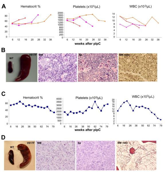
Figure 3. JAK2V617F mice develop PV and myelofibrosis.
(A) Blood parameters of mice with a PV-like phenotype displaying a marked increase in hematocrit and a fall in their platelet counts. (B) Mice with a PV-like phenotype show splenomegaly; BM hematoxylin and eosin (BM) showing erythroid and megakaryocytic hyperplasia with clustering and highly pleomorphic morphology; spleen hematoxylin and eosin (Sp) showing megakaryocytic and erythroid hyperplasia; reticulin stain showing no fibrosis in BM (BM retic) and spleen (not shown). (C) Blood parameters of a mouse with BM fibrosis displaying a gradual decline of blood count parameters, including hematocrit and white cells, and an increase in its platelet counts. (D) Splenomegaly in mouse with BM fibrosis; BM hematoxylin and eosin (BM) showing granulocytic hyperplasia with reduced megakaryocytic and erythroid cells; spleen hematoxylin and eosin (Sp) showing megakaryocytic, erythroid, and granulocytic expansion; reticulin stain showing fibrosis in BM (BM retic) but not in spleen (not shown).

