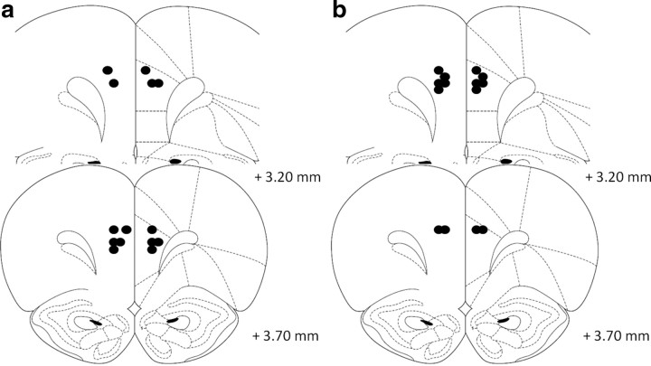
Figure 4.
Schematic representation of the position of the injector tips in experiment 3 as revealed by histological analysis. Guanfacine (a) (0.005 μg/0.5 μl per side) and α-flupenthixol (b) (15 μg/0.5 μl per side) were microinfused into the dPL (n = 7 for both). Drawings adapted from Paxinos and Watson (1998).

