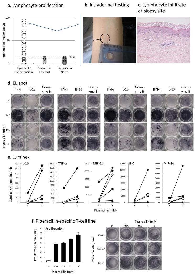
Figure 4. Piperacillin-specific stimulation of peripheral blood mononuclear cells and T-cell lines from hypersensitive patients.
(A) PBMC from 19 hypersensitive patients were specifically stimulated with piperacillin (SI above 2). (B) Positive intradermal skin test from 1 of 4 patients presenting with cutaneous signs and a strong in vitro proliferative response against piperacillin. (C) A biopsy of the maculopapular reaction site. Immune-stain confirmed the lymphocytic infiltrate as being almost entirely T-cell in character, with a CD3+, CD45RO+ phenotype. Both CD4+ and CD8+ subsets were present. (D) Piperacillin-specific-specific IFN-γ, IL13 and granzyme B ELIspot. The figure shows PBMC from 4 hypersensitive patients stimulated with piperacillin for 48 h. (E) Multiplex analysis of cytokines/chemokines secreted from hypersensitive patient PBMC (n=5) incubated with stimulatory concentrations of piperacillin. (F) Concentration-dependent proliferation and IFNγ secretion by a piperacillin-responsive T-cell line. T-cell lines were generated by repetitive stimulation of blood lymphocytes with piperacillin and irradiated autologous PBMC in IL-2 containing medium.

