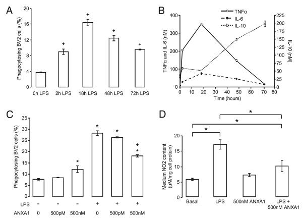
FIGURE 5.
Inflammatory activation of BV2 cells significantly alters the characteristics of their phagocytosis of PC12 cells, a change that can be restored by application of pharmacological concentrations of ANXA1. A, Preincubation of BV2 cells with 50 ng/ml LPS significantly enhances the phagocytosis of vehicle-treated PC12 cells in a time-dependent manner; data are mean ± SEM, n = 3. *p < 0.05 versus phagocytosis of PC12V in control conditions. B, BV2 cells were incubated for 0, 2, 6, 18, 48, and 72 h with 50 ng/ml LPS and then cocultured with PC12V for 2 h. Culture medium content of TNF-α, IL-6, and IL-10 were then measured by ELISA; data are mean ± SEM, n = 3; for all time points, levels of all three cytokines are significantly greater than control with p < 0.05. C, High (500 nM) but not low (500 pM) ANXA1 reverses the effect of LPS (18 h preincubation, 50 ng/ml) upon BV2 cell phagocytosis of vehicle-treated PC12 cells; data are mean ± SEM, n = 3. *p < 0.05 versus untreated BV2 cells; +p < 0.05 versus LPS-treated BV2 cells. D, Treatment of BV2 cells for 18 h with LPS induces significant accumulation of NO2−, an effect blocked by treatment with 500 nM human rANXA1 (data are mean ± SEM, n = 3). *p < 0.05).

